-
- Revolutionizing Healthcare AI: Unleashing AI Medical Imaging for Unmatched Diagnostic Accuracy
- Understanding AI in Medical Imaging
- Configuration Steps for Implementing AI in Medical Imaging
- Step 1: Define Objectives
- Step 2: Data Collection
- Step 3: Choose the Right AI Framework
- Step 4: Model Training
- Step 5: Model Evaluation
- Step 6: Deployment
- Practical Examples of AI in Medical Imaging
- Example 1: Stanford University Medical Center
- Example 2: Zebra Medical Vision
- Best Practices for AI Medical Imaging
- Case Studies and Statistics
- Conclusion
Revolutionizing Healthcare AI: Unleashing AI Medical Imaging for Unmatched Diagnostic Accuracy
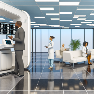
The integration of artificial intelligence (AI) in healthcare, particularly in medical imaging, is transforming the landscape of diagnostics. With the ability to analyze vast amounts of data quickly and accurately, AI is enhancing the precision of diagnoses, reducing human error, and ultimately improving patient outcomes. This guide delves into the configuration steps, practical examples, best practices, and case studies that illustrate how AI is revolutionizing medical imaging.
Understanding AI in Medical Imaging
AI in medical imaging involves the use of machine learning algorithms and deep learning techniques to interpret medical images such as X-rays, MRIs, and CT scans. These technologies can identify patterns and anomalies that may be missed by the human eye, leading to earlier and more accurate diagnoses.
Configuration Steps for Implementing AI in Medical Imaging
To successfully implement AI in medical imaging, follow these actionable steps:
Step 1: Define Objectives
- Identify specific diagnostic challenges you aim to address.
- Set measurable goals for accuracy and efficiency improvements.
Step 2: Data Collection
- Gather a diverse dataset of medical images relevant to your objectives.
- Ensure data is labeled accurately by qualified radiologists.
Step 3: Choose the Right AI Framework
- Select a machine learning framework such as TensorFlow or PyTorch.
- Consider pre-trained models for faster implementation.
Step 4: Model Training
Utilize the following code snippet to train your model:
import tensorflow as tf
from tensorflow.keras import layers, models

# Load and preprocess data
train_images, train_labels = load_data()

# Build the model
model = models.Sequential()
model.add(layers.Conv2D(32, (3, 3), activation='relu', input_shape=(image_height, image_width, 1)))
model.add(layers.MaxPooling2D((2, 2)))
model.add(layers.Flatten())
model.add(layers.Dense(64, activation='relu'))
model.add(layers.Dense(num_classes, activation='softmax'))

# Compile the model
model.compile(optimizer='adam', loss='sparse_categorical_crossentropy', metrics=['accuracy'])

# Train the model
model.fit(train_images, train_labels, epochs=10)
Step 5: Model Evaluation
- Use a separate validation dataset to evaluate model performance.
- Adjust hyperparameters based on evaluation results.
Step 6: Deployment
- Integrate the AI model into existing imaging systems.
- Ensure compliance with healthcare regulations and data privacy laws.
Practical Examples of AI in Medical Imaging
Several healthcare institutions have successfully implemented AI in their imaging departments:
Example 1: Stanford University Medical Center
Stanford developed an AI algorithm that can detect pneumonia in chest X-rays with an accuracy comparable to radiologists. This system has been instrumental in triaging patients and prioritizing cases based on urgency.
Example 2: Zebra Medical Vision
Zebra Medical Vision offers AI solutions that analyze medical imaging data to identify various conditions, including cardiovascular diseases and cancers. Their algorithms have been validated against large datasets, demonstrating high accuracy rates.
Best Practices for AI Medical Imaging
To maximize the effectiveness of AI in medical imaging, consider the following best practices:
- Regularly update and retrain models with new data to maintain accuracy.
- Involve radiologists in the development process to ensure clinical relevance.
- Implement robust data governance policies to protect patient information.
Case Studies and Statistics
Research indicates that AI can improve diagnostic accuracy significantly:
- A study published in Nature found that AI algorithms outperformed radiologists in detecting breast cancer in mammograms by 9.4%.
- According to a report by Accenture, AI applications in healthcare could save the industry $150 billion annually by 2026.
Conclusion
The integration of AI in medical imaging is not just a trend; it is a revolution that promises to enhance diagnostic accuracy and improve patient care. By following the outlined configuration steps, leveraging practical examples, adhering to best practices, and considering the supporting statistics, healthcare providers can effectively implement AI solutions that lead to better outcomes. As technology continues to evolve, staying informed and adaptable will be key to harnessing the full potential of AI in healthcare.